Surgical Management Of Subserosal (Interstitial) Urinary Bladder Rupture In Association With Pyometra In A 14 year old Lhasa Apso Dog.
Introduction:
Pyometra is an infection of the uterus with the accumulation of pus material, and is by far the most common disease of the uterus in an intact bitch. Pyometra generally occurs in middle-aged dogs of around 7–8 years of age.
Sub serosal (Interstitial) bladder rupture, is a very rare incidental type, compared to other Urinary bladder rupture classifications. It happens mostly due to blunt pressurizing trauma caused mostly by organ compressions, then any external injuries.
Case History and Clinical Examination
A14 yrs old Adult, intact female, Lhasa Apso, was presented to the Sanchu Veterinary hospital, Palavakkam ECR.
- With a history of sudden belly enlargement happening since 4 to 5 days.
- The caretakers said the dog is drinking more water quite often than eating food.
- Physical examination, the abdomen was tender and hard on palpation, the dog was unable to bear weight and walk. Abdominal palpation, intestinal loops were normal, bladder was intact, tenderness was felt.
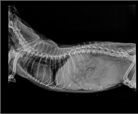
- Suggested the caretaker for X ray, (Fig. A) there is complete uterine enlargement pushing the intestines and spleen. Rectum was filled with faecal matter.
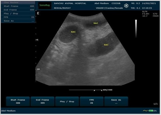
- Ultrasonography of the uterus shows multiple anechoic sacculation with normal viscera (Fig B) and diagnosis of Canine pyometra.
Clinical parameters:
HR 110bpm, RR 56 per min, TEMP 102.9°F, LN Swollen, CMM pale pink CRT 1sec, Hydration status 1 %
Surgical treatment and Post-operative care:
Since the dog was not having an appetite so it was kept on fluids one day before surgery (The intravenous cannula is fixed).
Inj RL 60ml IV, Inj Metro 60mg IV slowly, Inj Ceftri+tazo 150mg IV slowly, Inj Dicyclomine 6mg IV, Inj Panto 12mg IV.
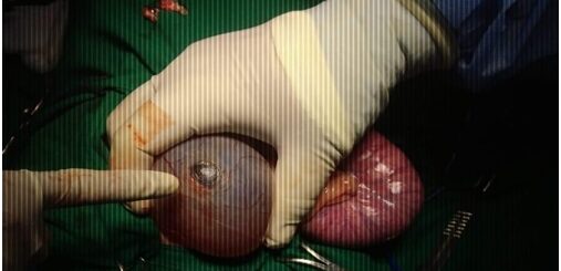
Under general anesthesia (isoflurane @ 3 MAC throughout the surgery), the animal was positioned in the ventrodorsal recumbency, the surgical site was clipped and prepared aseptically with Iodine scrub and Chlorhexidine solution. An abdominal midline celiotomy was performed. The infected uterus was isolated, which was compressing the bladder. While evacuating the bladder, the serosa of the bladder was ruptured exposing the mucosa (Fig D). There were no serosal adhesions between the ruptured portion to the uterus. The pus accumulated uterus was removed by Ovariohysterectomy procedure (Fig D), followed with Millers Knots on both ovaries and uterine blood vessels.
The mucosa of the bladder was intact, with no perforation or leaks or inflammation. The serosal surfaces of the rupture were trimmed so as to obtain clean, smooth, and regular wound edges. Six simple interrupted sutures were done to appose the serosa of the bladder, using Monocryl (polydioxanone) 4-0. The bladder was omentalized and fixed along with the omentum for faster healing (Fig E). The surgical site was lavage with warm Normal Saline and Metronidazole, checked for any signs of peritonitis (no peritoneal infection), urine leak, bleeding, and then covered the abdominal viscera with omentum. The abdominal muscles, subcutaneous tissue, and skin were closed using PGA (polyglycolic acid) 1-0. Postoperatively the dog was admitted for 3 days for fluid therapy and received systemic antibiotics along with analgesics. The dog started to eat on the 2nd day of post-operative care and was fed with A/d Hills commercial diet. The dog recovered uneventfully without any peritonitis, renal insufficiency, urinary tract infection, and inconvenience (Fig. F).
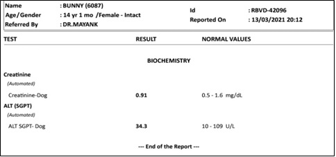
Fig C. Blood test reports indicating the dog is mildly anemic and thrombocytopenic 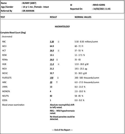
Fig C. Blood test reports indicating the dog is mildly anemic and thrombocytopenic 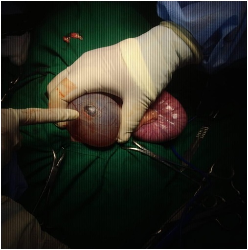
Fig D Sub Serosal bladder rupture and Ovariohysterectomy procedure of the pyometra. 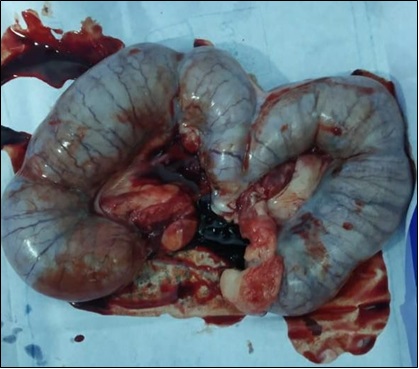
Discussion:
Pyometra is defined as infection and inflammation of the uterine glandular tissue with the accumulation of purulent material in an intact bitch. The risk of developing the condition within 10 years of age is estimated to be 23–24%. Traditionally, pyometra has been considered the end stage of cystic endometrial hyperplasia, which occurs due to an abnormal uterine response to progesterone. The uterine glandular tissue becomes edematous, thickened and cystic. Since progesterone inhibits myometrial contractility, the pus accumulates and this abnormal environment allows bacterial colonization. Pyometra generally occurs in middle-aged dogs of around 7–8 years of age, and is most commonly seen 4–8 weeks after estrus, although it can develop up to 4 months post-estrus. The condition is seen less commonly in the cat, and generally develops 1–4 weeks after estrous. Pyometra may occur in younger animals that have been given estrogen (mis-mating shots) or progestins for estrus suppression.
The organism most commonly cultured from pyometra is Escherichia coli, which is usually faecal in origin. Less frequently reported bacteria include Streptococcus, Pasteurella, Proteus and Klebsiella amongst many others. The route of infection is thought to be haematogenous and ascending. Pyometra is associated with cystitis in up to 70% of cases and this is thought likely to be as a result of purulent uterine material draining past the urethral orifice. Systemic effects are also encountered with pyometra. These include endotoxemia, renal insufficiency (due to immune complex glomerulonephritis or bacterial endotoxins, both of which are transient with successful treatment), immunosuppression (inhibition of lymphocyte activity, also transient) and systemic inflammatory response syndrome (SIRS). Diagnosis can be done by doing ultrasonography and x-ray followed by blood tests results to compare the differential.
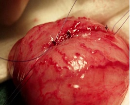
Fig E Bladder rupture repair and Omentalized 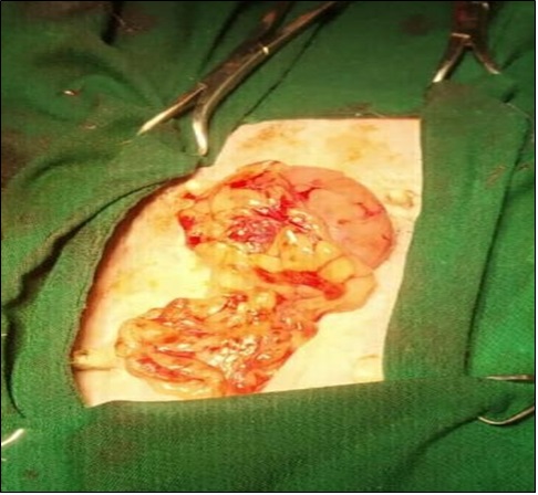
Fig E Bladder rupture repair and Omentalized
Urinary bladder rupture is pretty common in small animal practices since the trauma is a factor for most of it. Unlikely like in humans, there are 5 types of bladder rupture, namely,
- Intra-peritoneum
- Extra-peritoneum
- Bladder wall contusion
- Interstitial (sub serosal) and
- Combined peritoneum.
Classifications
- Intra-peritoneum: This injury is typically the result of a direct blow to the already distended bladder. The sudden increase in intravesical pressure causes intraperitoneal rupture of the bladder dome. CT cystography demonstrates intraperitoneal contrast material around bowel loops, between mesenteric folds, and in the paracolic gutters
- Extra-peritoneum: It is usually caused by penetrating trauma; the presumed mechanism is direct laceration of the bladder by bone fragments from a pelvic fracture. Extravasation is confined to the perivesical space in simple
extraperitoneal ruptures, whereas in complex extraperitoneal ruptures, contrast material extends beyond the perivesical space and may dissect into a variety of fascial planes and spaces. A thorough understanding of abdominopelvic anatomy and associated fascial planes will help avoid misdiagnosis of complex extraperitoneal rupture.
- Bladder Contusion: Bladder contusion is defined as an incomplete or partial tear of the bladder mucosa. Bladder contusion is believed to be the most common bladder injury in multi-trauma patients but is not in itself considered to be a major bladder injury.
- Sub serosal bladder rupture: Interstitial bladder injury is rare and is defined as an intramural or partial-thickness laceration intact mucosa. Consequently, CT cystography may demonstrate elliptical extravasation of contrast around the bladder.
- Combined peritoneum: Combined bladder rupture consists of simultaneous intraperitoneal and extraperitoneal injury. The prevalence of combined bladder rupture is 5%–12% in published series that include both penetrating and blunt trauma. CT cystography usually demonstrates extravasation patterns that are typical for both types of injury.
The most common types are the former 3, and very rarely subserosal bladder rupture is seen in animals. The cause factor for the occurrence of such bladder rupture is not knowledgeable. However, studies say, subserosal bladder rupture can occur due to constant stress or pressure on the serosal wall of the bladder caused by an organ or septic peritonitis or blunt pressurizing trauma or inflammatory reactions. Research is often going on to find out the cause. Diagnostic steps include FAST-ultrasonography and CT-scan based on the history. CT scan is an ideal method for bladder rupture diagnosis.
Conclusion: Sub serosal bladder rupture is not a very common type and not very popular in diagnostic experience except with CT Scan. It can be caused by this pyometra compression on it. The rupture was repaired showing no leaks and the dog recovered uneventfully.