Introduction:
Protozoa are classified under subkingdom protozoa. Protozoa are unicellular organisms having a distinct nucleus. The extranuclear part is cytoplasm which is further differentiated into inner endoplasm containing vacuoles, granules, and pigments. It contains the nucleus and cytoplasmic essential portion of protoplasm and its nutrition, either holophytic or phagotrophic type. It can be reproducing by asexual such as binary fission, budding and multiple fission, or sexual like syngamy, conjugation. Antoni van leeuwenhoek invented protozoa for the first time.
Notable protozoa found in the dog:
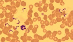
Trypanosoma evansi: Commonly cause a disease known as Surra. In Hindi Surra means rotten. It is most common in exotic breeds of dogs. Vector for transmission is flies such as tabanus, stomoxys. Antigen-antibody complex adhered to RBC and subjected to damage of RBC.
Clinical signs:
- It is marked by fever, anorexia, swelling of the head and throat.
- Swelling of the larynx, changes in the voice of the dog may occur.
- Inflammation of the conjunctiva, nasal discharge, blood in urine is observed.
- Skin seems smooth and red elevated papules in an infected animal.
Diagnosis
- Direct examination in fresh blood is the surest way of diagnosing acute infection.
- Chemical tests include
- Mercuric chloride test
- Formol gel test
- Stilbamidine test
- Jone’s nitric acid test
- Thymol turbidity test
- Blood smear examination for identification of the organism.
Prevention and control
- Bloodsucking flies act as vectors hence insecticide can be used for their control.
- Destruction of game animals which act as a reservoir of trypanosome.
- Treatment of infected animals as soon as possible.
- Quarantine of newly introduced animals and provide better management.
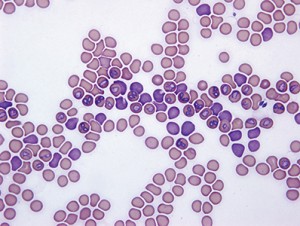
Mycoplasma haemocanis (Formerly called as Haemobartonella canis): It is found in the blood of an infected dog. Transmitted by direct transfer of infected blood or via arthropods vectors. Vertical transmission is also observed among dogs.
Clinical signs
- It causes the destruction of red blood cells hence, causing hemolytic anemia
- Acute hemolysis in dogs that have already damaged spleen.
- Lethargy, anorexia, fever and enlargement of the spleen occur
Diagnosis
- Diagnosis can be done based on Wright stained blood smear in which they appear as basophilic, round-shaped, in chains structure present on erythrocyte.
Prevention and control
- Control of arthropod vectors by using various available insecticides.
- Proper cleaning of the dog in order to avoid ticks.
- Removal of ticks from dogs either manually or with a suitable acaricide.
- Treatment should be done on an infected animal.
- Isolate the sick animal from a healthy herd to stop further transmission.
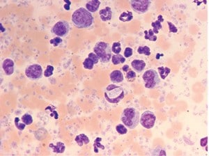
Hepatozoon canis: It causes old-world hepatozoonosis. The protozoan is transmitted by the brown dog tick, Rhipicephalussanguineus. It is transmitted by ingestion of ticks by dogs.
Clinical signs
- Polygranulomatous inflammation in tissues is seen.
- Fever, depression, weight loss, soreness, mucus discharge in the eyes is seen in an infected animal.
- Cervical, joint, or generalized pain is observed in affected dogs.
- Many dogs will not move to eat because of intense pain in the intestine.
Diagnosis
- Typically the number of neutrophils increased.
- A profound decrease in glucose level is observed.
- Microscopic examination of stained blood films to visualize parasite containing leukocytes.
Prevention and control
- Preventing the access of ticks in dog skin.
- Avoid ingestion of ticks by dogs
- Proper cleaning of kennel with detergent or warm water.
- Regular bath and cleaning of dog to avoid ticks.
- Treatment of infected animals with proper medications.
Sarcocystis cruzi: This protozoon is cosmopolitan in distribution. Oocysts are globular and subglobular. Sporulation occurs in the host. Sporulated oocyst contains 4 sporozoites. Globular organism found in immature Sarcocystis called metrocyst.
Clinical signs
- A high rise in temperature in the animal body is commonly observed.
- A pregnant dog may abort if it gets infected.
- Lameness, abnormal respiratory problems in dogs are observed.
- The sporozoites enter into the intestinal wall and further into the mesenteric lymph node
- Sporozoites initiate schizogony and schizonts are formed in the kidney and brain
- It causes enteritis, myositis, and nervous disorder
Diagnosis
- Examination of the fecal sample can be done for the detection of the oocyst.
- Post mortem examination of skeletal muscle, intestine can help in diagnosis.
Prevention and control
- Dogs should not be allowed to eat raw meat or dead animals
- Supplies of grain, feed should be properly covered.
- Treatment of infected dogs with proper medicine.
- Quarantine and provide good management of infected dogs.
Isospora burrowsi: These are found in dogs. Oocysts are spherical to ellipsoidal in shape. It occurs in the posterior small intestine and caecum of the dog. Laboratory mice and rats are transport hosts.
Isospora canis: These are found in the small intestine and also in the large intestine of the dog. House mice, hamsters act as a transport hosts. The oocytes are ellipsoidal or ovoid in shape.
Clinical signs
- Blood may be passed in faeces along with the heavy discharge of oocyst.
- Oocyst shed in faeces.
- Weight loss and dehydration due to diarrhea may be seen.
Diagnosis
- It is based on clinical signs like the presence of oocyst in faeces.
- During post mortem examination, a large number of developmental stages were found.
Prevention and control
- Isolation of infected animal from a healthy animal
- Proper sanitation of kennel with detergent
- Avoid raw meat, provide cooked meat.
- Fecal contamination to feed and water should be avoided.
- Proper control of insects with the help of many insecticides.
Babesia canis: These are found in the RBC of dogs. It is transmitted by the brown dog tick. A tick carrying B.canis sporozoites attaches to dogs and release sporozoites into the bloodstream. It can transmit through the placenta.
Clinical signs
- Lameness, weakness and vomiting is observed in an infected dog
- Pale mucous membrane and coffee-colored urine is the characteristic sign.
- Enlargement of spleen, jaundice, and anemia is observed in affected dogs.
Diagnosis
- It is based on clinical signs such as dark color urine discharge.
- It is not easy to find on blood smear so try to find on the capillary source rather than a blood vessel.
Prevention and control
- Treatment of affected animals with proper medications
- Control of ticks is important by proper cleaning of dogs.
- Isolation of infected animal from a healthy animal.
- Proper sanitation should be followed in the kennel
- Avoid raw, undercooked flesh.
Ehrlichia canis: It causes canine ehrlichiosis in dog. It is carried by ticks. It infects mostly monocytes in the blood. It can be transmitted by ticks like brown dog ticks.
Clinical signs
- It causes fever, widespread swelling of lymph nodes, and enlargement of the spleen.
- Loss of stamina and appetite observed
- Enlargement of the spleen and a decrease in the number of platelets are seen.
- Kidney failure, inflammation of eyes, lungs, and brain in long-term infection may be shown.
Diagnosis
- It is based on a blood smear examination.
- During post mortem examination lesions and changes were found in organs.
Prevention and control
- Control of ticks is very important either by removing them manually.
- Pull the ticks straight away make sure not to grasp or squeeze its body.
- Treatment of infected animals and proper sanitation of kennel should be done.
Balantidium coli: It is also known as ciliary dysentery. Its definitive host can be a dog, human, cow. Cysts are responsible for transmission through ingestion of infected food or water. Single trophozoite forms from each cyst.
Clinical signs
- Most cases are asymptomatic.
- Diarrhea with profuse blood and mucus may be seen.
- Nausea, vomiting, abdominal pain, and loss of appetite should be observed.
Diagnosis
- It can be detected in stool examination by their rotator motility, large kidney-shaped macronucleus.
- Through H&E stain cluster of trophozoites, cyst found in mucosa.
Prevention and control
- Treatment of carriers shedding the cysts.
- Hygienic rearing of pigs and prevention of pig to dog contact.
- Avoid contamination of food as well as giving raw meat to dogs.
- Provide quality and clean water to dogs.
- Infected dogs should quarantine from healthy animals.
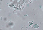
Giardia canis: It causes giardiasis cause of intestinal infection. It is mainly transmitted by eating cyst-infected food. Encystations occur as parasites transit towards the colon. Transmission mainly through the fecal-oral route.
Clinical signs
- It usually causes diarrhea, bloating, abdominal pain, and vomiting.
- The animal becomes weak and infirm in chronic conditions.
- It decreases the absorption in the intestine due to the shortening of microvilli.
Diagnosis
- It is diagnosed by the identification of cysts in feces using direct mounts.
- By fecal examination, the oval-shaped cyst may be seen.
Prevention and control
- Control of faeces as soon as possible disposing of potentially infectious waste.
- The cyst can be inactivated by quaternary ammonium compound, steam, or boiling water.
- Prompt and frequent removal of faeces.
- Proper cleaning of kennel with boiling water or detergent.
Entamoeba histolytica: It causes amoebiosis in dogs which causes persistent diarrhoea. Dogs are generally infected by eating infected food and water with the cyst. The affected dog shows acute or chronic colitis.
Clinical signs
- It usually causes diarrhoea, weight loss, and anorexia
- It mainly causes frequent diarrhoea in dogs.
Diagnosis
- It is diagnosed by the identification of cysts in faeces.
- Colonoscopy with a scraping of ulcerations is more effective.
- The cyst is typically seen in wet mounts while trophozoite is directly seen in saline smear.
Prevention and control
- Control of faeces as soon as possible disposing of potentially infectious waste.
- The cyst can be inactivated by the use of quaternary ammonium compound, steam, and boiling water.
- Frequent removal of faeces so that helps in removing the cyst.
- Isolation of infected animal from a healthy animal.
- Faecal contamination of feed and water should be avoided.
- Proper control of insects with the help of suitable insecticide.
*image credits Todays Veterinary Practice