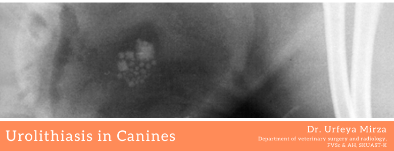
Urolithiasis is the process of formation of stones in the kidney, bladder, and/or urethra (urinary tract). The development of the stones is related to decreased urine volume or increased excretion of stone-forming components such as calcium, oxalate, urate, cystine, xanthine, and phosphate. The stones form in the urine collecting area (the pelvis) of the kidney and may range in size from tiny to staghorn stones the size of the renal pelvis itself.
Urolithiasis is a common health concern of multifactorial origin. This increase is largely attributable to changes in diet and lifestyle, leading to obesity. The incidence of urolithiasis has increased not only in the human population, but also in many other animals, affecting a wide range of species, including wild animals, farm animals, primarily ruminants and companion animals, largely dogs and cats. Risk factors that lead to the increase in the incidence of urolithiasis in nonhuman members of the animal kingdom seem to parallel that in humans. A number of investigators have developed models, primarily in rats and pigs, that somewhat mimic human nephrolithiasis. An accurate and reliable animal model may allow us to develop newer treatment algorithms and medications that may help to better understand the pathogenesis of stone formation and direct improved methods of stone prevention. We reviewed the incidence of stone disease in animals, describe the currently used animal models of nephrolithiasis and document the surgical and medical treatment of animal stone disease. The incidence of urolithiasis has not been established for the animal kingdom, although dogs, cats, and horses are the most common mammals to receive treatment for stone dis ease. Other animals, including non-mammals and mammals, have also been reported to form renal and bladder calculi, including pigs, sheep, birds, turtles, cattle, goats, whales and deer. The table provides an overview of various animal species affected by urinary tract stone disease. Most information on animal urolithiasis comes from the canine and feline literature. The incidence of urolithiasis in dogs and cats is believed to be between 0.2% and 3.0% (Bartges et al., 2004). Of the varying stone types magnesium ammonium phosphate hexahydrate (struvite) and calcium oxalate are the most common compositions in each species with calcium oxalate present in 55% of feline and 46% of canine lower urinary tract stones, and in 90% of feline upper tract stones. Traditional urological discussions of stones in canines involve urate stones in Dalmatian dogs. However, stones have been identified in more than 26 breeds of dogs. The implication of this high stone prevalence suggests a potential resource of animal subjects to better understand human stone disease. Even the changing epidemiological pattern in the struvite and calcium oxalate prevalence in canines is an interesting phenomenon that suggests dietary influences on stone disease and which ultimately may help us understand dietary factors in human stone disease (Stevenson et al., 2004).
Calculous formation involves a complex sequence of events, including the state of urinary saturation of calculogenic materials, and the presence of urinary inhibitors of crystallization, and various urinary promoters of crystal aggregation and growth (Bartges et al., 2004). To better understand the process of calculous formation and its promoting factors various animal models ranging from rats to dogs have been used, including diet induced urolithiasis; drug induced urolithiasis and genetically altered animals that form stones. Diet induced urolithiasis has been investigated for more than a quarter of a century.
Types of Stones:
Uric acid stones: Although they can form other types of calculi, Dalmatian dogs are traditionally recognized as being uniquely predisposed to uric acid calculi because of heritable defects in uric acid metabolism. There are 2 potential mechanisms promoting the formation of uric acid calculi in Dalmatians. The most significant one quantitatively appears to be that the liver does not oxidize available uric acid completely despite containing an adequate concentration of uricase. To a lesser extent the proximal renal tubules in Dalmatians reabsorb less uric acid than those in non-Dalmatians (Osborne et al., 1989). Little information is known about the mechanism of development of uric acid calculi in non-Dalmatian dogs. Potential predisposing factors are:
- increased renal excretion and urine concentration of uric acid,
- increased renal excretion, renal production or microbial urease production of ammonium ions,
- low urine pH and
- promoters or absent inhibitors of uric acid calculi formation.
Calcium oxalate stones: Excessive urinary excretion of calcium is a significant risk factor for calcium oxalate stone formation in humans, dogs and cats. As in humans, in dogs gastrointestinal hyper-absorption appears to be the most common reason for hyper-calciuria, followed by impaired renal absorption of calcium (renal leakage). In cats the consumption of diets supplemented with the urinary acidifier ammonium chloride is associated with increased urinary calcium excretion, which is analogous to human consumption of a high protein diet.4 Acidosis is known to increase urinary calcium excretion. Calcium ions and oxalate, which are the metabolic end product of ascorbic acid and several amino acids, complex to form a relatively insoluble salt, leading to increased urinary excretion of these compounds that may prompt calcium oxalate formation. Hyperoxaluria has been observed in cats consuming diets deficient in vitamin B6 and in those with decreased quantities of hepatic D-glycerate dehydrogenase. Hyperoxaluria has not been identified in dogs (Bartges et al., 2004).
Calcium Phosphate Stones: Calculi Calcium phosphate stones in animals are relatively uncommon and their underlying metabolic explanations are not as well understood as in humans. Renal tubular acidosis in dogs produces struvite stones. Primary hyperparathyroidism, hypercalcemic states and normocalcemic hypercalciuria are associated with brushite stone formation. Addressing the underlying cause is the main primary treatment and sometimes surgery is indicated if symptoms persist.
Surgical treatment
Surgical treatment of calculi should be reserved for animals with outflow obstruction and functional impairment of the associated kidney, and when there is no effective dissolution medical therapy available, i.e., calcium oxalate stones. Surgery also is indicated in cases in which there is a contraindication to low protein, mineral restricted diets, increasing stone growth despite medical therapy, anatomical defects of the urogenital tract that predispose to recurrent urinary tract infections and poor medical compliance. As in treating stone disease in humans, the surgical procedure of choice is determined primarily by the likelihood of complete removal of the calculus based on stone size, location and configuration (Ross et al., 1999). Surgical interventions range from endoscopic procedures with laser lithotripsy to open stone extraction.
Management
Dietary management appears to be the predominant therapy for canines and felines; medical therapy protocols aimed at arresting further calculi growth, dissolution and prevention have made a significant impact on recurrent urolithiasis. However, medical therapy is not always the most feasible treatment option. For animals in which medical therapy fails and that have symptomatic calculus surgical intervention is the option of choice. However, the morbidity and mortality of open surgery are an area of concern. Until minimally invasive technology becomes more widespread surgical stone removal still poses challenge to veterinary urology. Equipment costs, miniaturized endoscopes and adequate training are obstacles that veterinary medicine must address, just as human urology has faced in the past.